Avicenna Journal of Medical Biochemistry. 8(2):128-132.
doi: 10.34172/ajmb.2020.18
Brief Report
Comparison of Serum Iron, Zinc, and Selenium Levels in Premenopausal and Postmenopausal Women in Ekpoma, Nigeria: A Descriptive Study
Oloruntoba O. Festus 1  , Solomon O. Agbebaku 2, Blessing O. Idonije 3, Olarewaju M. Oluba 4, *
Author information:
1Department of Medical Laboratory Science, College of Medicine, Ambrose Alli University, Ekpoma, Nigeria
2Department of Chemical Pathology, College of Medicine, Ambrose Alli University, Ekpoma, Nigeria
3Department of Medical Biochemistry, College of Medicine, Ambrose Alli University, Ekpoma, Nigeria
4Department of Biochemistry, Food Safety & Toxicology Research Unit, College of Pure and Applied Sciences, Landmark University, Omu-Aran, Kwara State, Nigeria
*
Corresponding author: Olarewaju M. Oluba, Department of Biochemistry, Food Safety & Toxicology Research Unit, College of Pure and Applied Sciences, Landmark University, OmuAran, Kwara State, Nigeria. Tel: +2347030496639, Email:
oluba.olarewaju@lmu.edu.ng;
olubamike2000@yahoo.co.uk
Abstract
Background: Estrogen deficiency following menopause creates an imbalance in plasma micronutrient resulting in several degenerative pathological conditions, including hypertension, cardiovascular disease, osteoporosis, etc.
Objectives: The present study was designed to compare zinc (Zn), iron (Fe), and selenium (Se) concentrations between premenopausal and postmenopausal women.
Methods: In this descriptive study a total of 200 participants were classified into two groups of postmenopausal (age range: 46-75 years, served as experimental) and premenopausal (age range: 30-45 years, served as control). Each group consisted of 100 subjects. After obtaining informed consent from all participants, blood samples were collected from the antecubital fossa vein of each participant by venipuncture. The concentrations of Fe, Zn, and Se in each blood sample were determined using Atomic Absorption Spectrophotometer.
Results: No significant difference (P>0.05) was observed in serum Fe (114.24 ± 26.79 µg/dL), Zn (83.11 ± 20.45 µg/dL), and Se (41.99 ± 9.78 µg/dL) levels between the control and experimental groups. However, serum Fe and Zn showed progressive significant (P=0.04, 0.03, respectively) increase with increasing postmenopausal age. Conversely, serum Se concentration decreased significantly (P=0.03) with increasing menopausal age.
Conclusion: Although no significant difference was observed in serum levels of Fe, Zn, and Se between pre- and post-menopausal women, the progressive significant increase in the serum Fe and Zn levels as well as significant decrease in serum Se level with advancing post-menopausal age portend a great risk.
Keywords: Trace mineral, Menopause, Postmenopausal women
Copyright and License Information
© 2020 The Author(s); Published by Hamadan University of Medical Sciences.
This is an open-access article distributed under the terms of the Creative Commons Attribution License (
http://creativecommons.org/licenses/by/4.0), which permits unrestricted use, distribution, and reproduction in any medium provided the original work is properly cited.
Introduction
Menopause is an inevitable natural phenomenon accompanying aging in women. Attainment of menopause impacts several physiological and biochemical changes that most often impair the quality of life in women (1). Increased alterations or imbalance in nutrients including trace elements metabolism and vitamins are some of the attendant consequences of menopause. These biochemical disturbances accompanying menopause have been attributed to the decrease in estrogen level resulting from deranged lipid metabolism, insulin concentration, bone resorption (osteoporosis), cardiovascular diseases and related degenerative diseases, etc (2). Following the attainment of menopause, there is an augmented production of free radicals culminating in enhanced oxidative stress due to abrupt changes in hormonal status (3). Ultimately, endogenous antioxidant defense mechanism, both enzymatic and non-enzymatic, is suppressed in postmenopausal women in relation to premenopausal women (3). Enzymatic antioxidants such as glutathione peroxidase (GPx) and superoxide dismutase (SOD) are metallo-enzymes requiring utilizing trace metals as cofactors. GPx is a seleno-metalloenzyme while SOD uses zinc (Zn) and copper (Cu) as cofactors. The decrease in body antioxidant status due to hormonal imbalance in post-menopausal women has been observed to be directly associated with serum trace element level (4).
The progression in age following menopause is usually associated with certain physiologic alterations leading to profound changes in circulating levels of nutrient-binding proteins and micronutrients within the cell. Cases of increased incidences of nutrient imbalance resulting from trace elements and vitamins deficiencies in menopause have been reported (3). Zn is essential in regulating calcitonin release from thyroid gland and thus exerting a great influence on bone turnover (4). Iron (Fe), which is another important trace element affected by menopause, plays crucial roles in oxygen transport, electron transfer in oxidative phosphorylation, and regulation of cell growth and differentiation. It also acts as a catalyst in the generation of free radicals, which are most often injurious to the cells. High plasma ferritin (stored form of Fe) concentration above normal has also been implicated in the pathogenesis of ischemic heart disease (5). Selenium (Se), on the other hand, acts as a cofactor to GPx, an important endogenous antioxidant protein which defends the cells against free radical-mediated oxidative damage (6). Currently, there is limited data on serum trace metal level in postmenopausal women in Nigeria. Data on trace metal or micronutrient metabolism in postmenopausal women can contribute significantly in the monitoring and management of complications due to the onset of menopause in women. Accordingly, the present study was designed to compare serum levels of Fe, Zn, and Se between premenopausal and postmenopausal women. Moreover, it evaluated the changes in the concentration of these trace minerals with advancing post-menopausal age.
Materials and Methods
Study Area
This is a cross-sectional study conducted in Ekpoma, Edo State, Nigeria. Ekpoma is a town located at latitude 6.75°N and longitude 6.13°E having an estimated population of 59 618 people. The town has grown into an urban center after its designation as headquarters and as the host of Ambrose Alli public university. The inhabitants are mainly students, civil servants, artisans, and farmers.
Ethical Approval
Approval for the study was granted by the Research and Ethics Committee of Ambrose Alli University, Ekpoma, Edo State, Nigeria (Approval number: AAU/REC/HS1116).Questionnaires were administered to collect information on demographical and medical histories. Subjects with symptoms of respiratory distress, infection, having any form of malignant disease, and those under any special medical treatment were excluded. Subjects that met the inclusion criteria signed a written informed consent letter.
Sample Size
The study population consisted of 100 post-menopausal women (age range: 46-75 years) as experimental group and 100 premenopausal women (age range: 30-45 years) as control. Post-menopausal group consisted of women who had missed their menstrual period (amenorrhea) for at least one year and were not on estrogen therapy or any such supportive treatment for menopausal symptoms for at least 6 months prior to the study. In addition, all subjects had normal blood pressure and were not suffering from any known systemic or endemic disorders.
Sample Collection
This cross-sectional study was carried out within 6 months (March – September, 2016). Samples were collected three times a week (Mondays, Wednesdays, and Fridays). To collect samples,5 mL of blood samples were collected from the antecubital fossa vein of each participant by venipuncture in the morning before the commencement of daily activities. The samples were dispensed into plain sample bottles and left undisturbed for clotting and retraction. The blood samples were centrifuged at 3000 rpm for 10 minutes. The serum was separated into new, clean, and dry plain containers and stored frozen until analysis.
Sample Analysis
In each sample, Fe, Se, and Zn levels were estimated by Atomic Absorption Spectrophotometer (Chemtech Analytical, USA) using a hollow cathode lamp at 214.1 nm. The instrument was calibrated with Chem LabTM standard solution (National Bureau of Standards, Washington DC, USA). All analysis was carried out in the Clinical Chemistry Laboratory at University College Hospital, Ibadan, Oyo State, Nigeria.
Statistical Analysis
Data are presented as means ± standard error of the mean (SEM) of 100 determinations. Difference between means were compared using student’st test. P value < 0.05 was considered significant.
Results
Serum Fe, Zn, and Se levels in postmenopausal and premenopausal women are presented in Table 1. Although serum Fe concentration was higher in premenopausal group (120.79 ± 27.70 µg/dL) compared to postmenopausal one (114.24 ± 26.79 µg/dL), the difference in Fe level between the two groups was not statistically significant (P=0.89). Similarly, the mean serum Zn and Se levels were higher in premenopausal group (88.90 ± 20.22 µg/dL and 44.3 ± 10.2 µg/dL, respectively) compared to postmenopausal one (83.11 ± 20.45 µg/dL and 42.0 ± 9.8 µg/dL, respectively); however, the observed differences were not significant (P= 0.73).
Table 1.
Serum Fe, Zn, and Se Levels in Premenopausal and Postmenopausal Women
| Serum parameters |
Pre-menopausal
(n = 100) |
Post-menopausal
(n = 100) |
P Value |
Remark |
| Fe (µg/dL) |
120.8 ± 27.7 |
114.2 ± 26.8 |
0.89 |
P > 0.05 |
| Zn (µg/dL) |
88.9 ± 20.2 |
83.1 ± 20.5 |
0.73 |
P > 0.05 |
| Se (µg/dL) |
44.3 ± 10.2 |
42.0 ± 9.8 |
0.73 |
P > 0.05 |
Values are presented as means ± SEM.
Changes in serum Fe, Zn, and Se levels with respect to advancing postmenopausal age are presented in Table 2. Serum Fe concentration increased with age, except for the postmenopausal women in the age range of 56-60 years (97.92 ± 23.06 µg/dL), where a reduction was observed compared to those in the age range of 51-55 years (100.22 ± 18.72 µg/dL) (Figure 1A). The observed differences were statistically significant (P ≤ 0.04). Specifically, the mean serum Fe in postmenopausal women between the age ranges of 61-65 years (107.08 ± 20.92 µg/dL), 66-70 years (127.06 ± 22.85 µg/dL), and +71 years (134.41 ± 21.31 µg/dL) were significantly higher (P = 0.04) compared to those in the age range of 46-50 years (80.47 ± 7.96 µg/dL).
Table 2.
Serum Fe, Zn, and Se Levels in Relation to Postmenopausal Age
| Serum Level |
Postmenopausal Age (y) |
P Value |
Remark |
46-50
(n = 30) |
51-55
(n = 20) |
56-60
(n = 20) |
61-65
(n = 10) |
66-70
(n = 10) |
Above 70
(n = 10) |
| Fe (µg/dL) |
80.5 ±
8.0 |
100.2 ±
18.7 |
97.9 ±
23.1 |
107.1 ±
20.9* |
127.1 ±
22.9* |
134.4 ±
21.3* |
0.04 |
P < 0.05 |
| Zn (µg/dL) |
59.0 ±
5.8 |
71.8 ±
14.7 |
71.7 ±
16.9 |
76.01±
21.3* |
93.1 ±
16.7* |
98.2 ±
15.6* |
0.03 |
P < 0.05 |
| Se (µg/dL) |
49.5 ±
2.9* |
46.6 ±
8.4* |
41.6 ±
6.1* |
35.9 ±
8.4 |
35.9 ±
7.4 |
29.1 ±
7.8 |
0.03 |
P < 0.05 |
Values are presented as means ± SEM. * Statistically significant at P < 0.05 compared with 46-50 years.
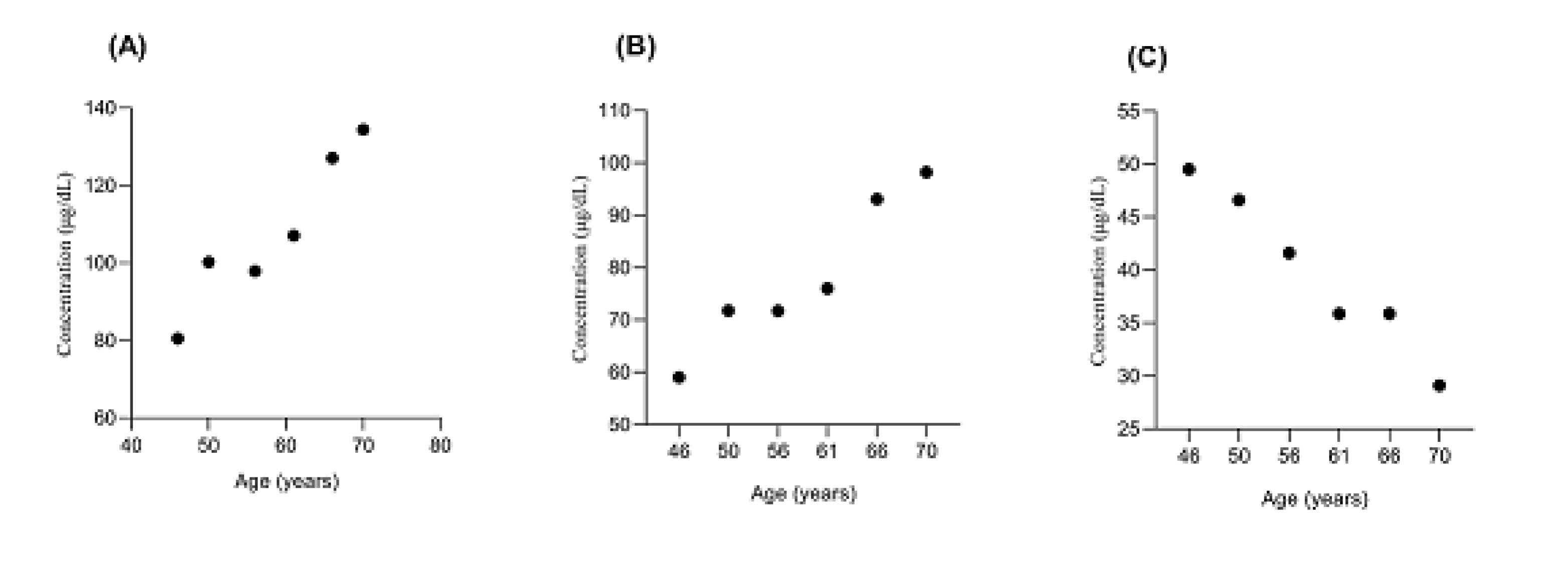
Figure 1.
Scattered Plots Showing the Dependence of Postmenopausal age with Serum (A) Fe, (B) Zn, and (C) Se Levels.
.
Scattered Plots Showing the Dependence of Postmenopausal age with Serum (A) Fe, (B) Zn, and (C) Se Levels.
Similarly, the mean serum Zn level increased as postmenopausal age progressed (Figure 1B). The increase in serum Zn level with advancing postmenopausal age was statistically significant (P = 0.04). The results showed that the mean serum Zn levels of postmenopausal women in the age ranges of 61-65 years (76.01 ± 21.28 µg/dL), 66-70 years (93.08 ± 16.74 µg/dL), and +71 years (98.24 ± 15.59 µg/dL) were significantly higher (P = 0.03) compared to postmenopausal women in the age range of 46-50 years (58.95 ± 5.83 µg/dL).
Conversely, serum Se level in postmenopausal women decreased progressively with advancing age (Figure 1C). The age- related decrease in the level of serum Se in postmenopausal women was statistically significant (P = 0.03). Specifically, there was a significantly (P = 0.03) higher serum Se level in postmenopausal women in the age ranges of 46-50 years (49.5 ± 2.9 µg/dL), 51-55 years (46.6 ± 8.4 µg/dL), and 56-60 years (41.6 ± 6.1 µg/dL) compared to postmenopausal women in the age ranges of 61-65 years (35.9 ± 8.4 µg/dL), 66-70 years (35.9 ± 7.4 µg/dL), and +70 years (29.1± 7.8 µg/dL).
Discussion
The inevitable process of menopause in women usually presents with its some metabolic consequences. Perturbation in cellular trace element concentrations is an associated factor accompanying the onset of menopause in women. Data obtained from this study showed similar Fe, Zn, and Se concentrations in postmenopausal and premenopausal women. This is in contradiction with earlier report of a decrease in serum Fe level in postmenopausal women from Sudan (7). Lynch et al (8) also demonstrated a decrease in Fe level in elderly women due to the aging process. The documented similarity in Fe, Zn, and Se levels between post- and pre-menopausal women in this study is consistent with the reports of Bureau et al (9) and Ansar et al(10), that showed a non-appreciable difference in trace elements concentrations between postmenopausal and premenopausal women. As mentioned, Fe is crucial in the transport of oxygen between tissues as well as in the transfer of electron in electron transport chain and in the regulation of cell growth and differentiation. In addition, it functions as a facilitator in free radical generation (11). Imbalance in cellular Fe level has been reported to cause serious health concerns.
Zn is an essential antioxidant in the human body where it serves as an important component of several enzymes involved in protecting the cells against oxidative assaults (3). In the current study, no significant difference in serum Se concentration was observed between postmenopausal and premenopausal women. This observation is inconsistent with the results of Manafa et al (12) who reported an increase in serum Se level in postmenopausal women. Moreover, Se is an important cofactor for the reduction of antioxidant enzyme such as GPx, an enzyme which scavenge potentially harmful oxidizing agent in the cell (6).
We observed that serum Fe increased with postmenopausal age, except for the postmenopausal women in the age range of 56-60 years, in which there was a reduction in Fe level. This observation is in agreement with the findings of Liu et al (4) who claimed that plasma ferritin concentrations usually increase with age until approximately 60 years where it reaches a plateau. Similarly, previous studies by Kato et al (13) and Zacharski et al (14) reported an increase in serum Fe concentration with increasing age in apparently healthy postmenopausal women. High serum Fe level in postmenopausal women could be a risk factor in the development of postmenopausal osteoporosis. Postmenopausal osteoporosis is one of the numerous associated health challenges with menopause-related estrogen deficiency (15), establishing a possible link between oxidative stress and menopause.
Similarly, in this study, serum Zn level increased with advancing age in postmenopausal women. In a study by Benes et al (16) involving a population of Czech women, plasma Zn concentration was demonstrated to increase with age. Increase serum Zn concentration in postmenopausal women could be an associated effect of enhanced bone resorption resulting from estrogen deficiency (4).
Our data also demonstrated an age-related decrease in serum Se level in postmenopausal women. This is in line with earlier reports of a declining Se concentration with age in postmenopausal women (17). Decreased Se levels observed in previous studies could be a reflection of inadequate dietary intake (18), several age-related physiological problems, low appetite, sedentary lifestyle, or a lack of physical activity (19). Moreover, the consequent elevated release of free radicals due to high serum Fe concentration, as observed in this study, may have led to a decline in Se concentration.
Conclusion
The results of this study did not show a significant difference in Fe, Zn, and Se levels between premenopausal and postmenopausal women. However, the progressive increase in the serum Fe and Zn and a decrease in serum Se concentrations with advancing postmenopausal age indicate a great risk with regard to estrogen deficiency. These results could be helpful in assessing the health status of postmenopausal women and improving the treatment methods. Identifying the magnitude of trace elements deficiencies among postmenopausal women is essential for evidence-based intervention modalities.
Conflict of Interest Disclosures
The authors declare that there is no conflict of interests.
References
- Kaur K, Gupta R, Saraf SA, Saraf SK. Zinc: The Metal of Life. Compr Rev Food Sci Food Saf 2014; 13(4):358-76. doi: 10.1111/1541-4337.12067 [Crossref] [ Google Scholar]
- Ansar S, Alhefdhi T, Aleem AM. Status of trace elements and antioxidants in premenopausal and postmenopausal phase of life: a comparative study. Int J Clin Exp Med 2015; 8(10):19486-90. [ Google Scholar]
- Vincent J, Inassi J. Comparison of oxidative stress between premenopausal and postmenopausal women. Natl J Physiol Pharm Pharmacol 2020; 10(5):359-62. [ Google Scholar]
- Liu JM, Hankinson SE, Stampfer MJ, Rifai N, Willett WC, Ma J. Body iron stores and their determinants in healthy postmenopausal US women. Am J Clin Nutr 2003; 78(6):1160-7. doi: 10.1093/ajcn/78.6.1160 [Crossref] [ Google Scholar]
- González-Estecha M, Palazón-Bru I, Bodas-Pinedo A, Trasobares E, Palazón-Bru A, Fuentes M. Relationship between serum selenium, sociodemographic variables, other trace elements and lipid profile in an adult Spanish population. J Trace Elem Med Biol 2017; 43:93-105. doi: 10.1016/j.jtemb.2016.12.002 [Crossref] [ Google Scholar]
- Wang N, Tan HY, Li S, Xu Y, Guo W, Feng Y. Supplementation of micronutrient selenium in metabolic diseases: its role as an antioxidant. Oxid Med Cell Longev 2017; 2017:7478523. doi: 10.1155/2017/7478523 [Crossref] [ Google Scholar]
- Yassein RB, Alseedig NO, Abdallah SK, Mohammed AA, Alballah NA, Syid MA. Hematological parameters among Sudanese patients with chronic renal failure. Int J Res Granthaalayah 2016; 4(16):50-4. [ Google Scholar]
- Lynch SR, Finch CA, Monsen ER, Cook JD. Iron status of elderly Americans. Am J Clin Nutr 1982; 36(5 Suppl):1032-45. doi: 10.1093/ajcn/36.5.1032 [Crossref] [ Google Scholar]
- Bureau I, Anderson RA, Arnaud J, Raysiguier Y, Favier AE, Roussel AM. Trace mineral status in post menopausal women: impact of hormonal replacement therapy. J Trace Elem Med Biol 2002; 16(1):9-13. doi: 10.1016/s0946-672x(02)80003-7 [Crossref] [ Google Scholar]
- Ansar S, Alhefdhi T, Aleem AM. Status of trace elements and antioxidants in premenopausal and postmenopausal phase of life: a comparative study. Int J Clin Exp Med 2015; 8(10):19486-90. [ Google Scholar]
- Qamar K, Saboor M, Qudsia F, Khosa SM, Moinuddin Moinuddin, Usman M. Malabsorption of iron as a cause of iron deficiency anemia in postmenopausal women. Pak J Med Sci 2015; 31(2):304-8. doi: 10.12669/pjms.312.6462 [Crossref] [ Google Scholar]
- Manafa PO, Nna CD, Chukwuma GO, Onyenekwe CC, Ihim AC, Iloghalu EU. Cobalt, copper, selenium and zinc levels in pre-menopausal and post-menopausal women in Nnewi, South-East Nigeria. Orient J Med 2015; 27(3-4):93-8. [ Google Scholar]
- Kato I, Dnistrian AM, Schwartz M, Toniolo P, Koenig K, Shore RE. Risk of iron overload among middle-aged women. Int J Vitam Nutr Res 2000; 70(3):119-25. doi: 10.1024/0300-9831.70.3.119 [Crossref] [ Google Scholar]
- Zacharski LR, Ornstein DL, Woloshin S, Schwartz LM. Association of age, sex, and race with body iron stores in adults: analysis of NHANES III data. Am Heart J 2000; 140(1):98-104. doi: 10.1067/mhj.2000.106646 [Crossref] [ Google Scholar]
- Bonaccorsi G, Piva I, Greco P, Cervellati C. Oxidative stress as a possible pathogenic cofactor of post-menopausal osteoporosis: existing evidence in support of the axis oestrogen deficiency-redox imbalance-bone loss. Indian J Med Res 2018; 147(4):341-51. doi: 10.4103/ijmr.IJMR_524_18 [Crossref] [ Google Scholar]
- Benes B, Spevácková V, Smíd J, Batáriová A, Cejchanová M, Zítková L. Effects of age, BMI, smoking and contraception on levels of Cu, Se and Zn in the blood of the population in the Czech Republic. Cent Eur J Public Health 2005; 13(4):202-7. [ Google Scholar]
- Bates CJ, Thane CW, Prentice A, Delves HT. Selenium status and its correlates in a British national diet and nutrition survey: people aged 65 years and over. J Trace Elem Med Biol 2002; 16(1):1-8. doi: 10.1016/s0946-672x(02)80002-5 [Crossref] [ Google Scholar]
- Schmuck A, Roussel AM, Arnaud J, Ducros V, Favier A, Franco A. Analyzed dietary intakes, plasma concentrations of zinc, copper, and selenium, and related antioxidant enzyme activities in hospitalized elderly women. J Am Coll Nutr 1996; 15(5):462-8. doi: 10.1080/07315724.1996.10718625 [Crossref] [ Google Scholar]
- Campbell D, Bunker VW, Thomas AJ, Clayton BE. Selenium and vitamin E status of healthy and institutionalized elderly subjects: analysis of plasma, erythrocytes and platelets. Br J Nutr 1989; 62(1):221-7. doi: 10.1079/bjn19890022 [Crossref] [ Google Scholar]